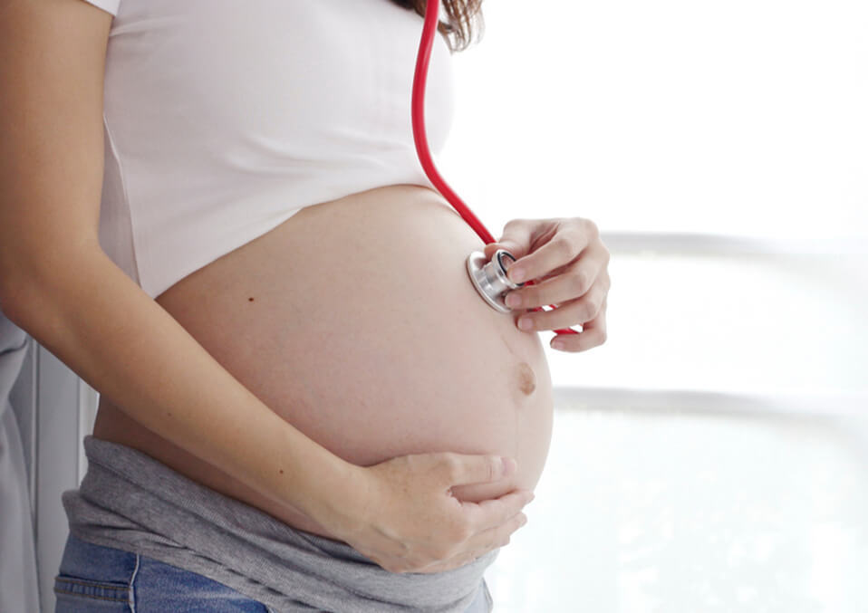
Having discovered that she is pregnant, she is probably looking forward to hearing your baby’s heartbeat, so how can you hear the baby’s heartbeat with a stethoscope?
It is one of the most wonderful sounds out there. Furthermore, it presumably sounds simply like a registration; yet consistently there are enormous changes in the heart and circulatory framework!
Your Child’s Mind Begins To Develop.
This sweet, sweet heartbeat at Week 4 has not yet arrived; but the different blood vessels will soon be in your baby’s heart and the circulatory system (blood) in the early stages forming in your embryo.
The heart will eventually form a heart and valves (from the heart to release blood to the body); which are twisted and separated into tubes.
In fact, at Week 5; the heart tube cannot hear it, but spontaneously; start to beat.
During these initial couple of weeks; vein antecedents likewise start to shape in the incipient organism.
When You First Hear The Heartbeat Of The Baby.

The heart of your baby hitting 110 times for 6 weeks and now has four hollow chambers; each with an inlet and outlet to allow blood flowing inside and outside each chamber; this number increases to 150-170 beats per minute.
It’s twice as quick as you!
With all this growth; you can hear your baby’s heartbeat for the first time around Week 9 or Week 10 of pregnancy.
Now it’s going to be about 170 beats/minute; the rate of decline from now on.
Your doctor or midwife will place a portable ultrasound machine; called a Doppler in your stomach to amplify the sounds you dictate.
Haven’t you heard? Don’t worry, it’s not just that your shy boyfriend or girlfriend is hiding in the corner of your womb or has a back (or next visit) after a few weeks.
The wonderful sound of your baby’s heartbeat will be heard for your hearing pleasure.
Your professional check-in is easy; convenient and OK; so please do not hesitate to contact us before you worry.
Ultrasound And Innate Heart Abandon.
Somewhere close to 6 and 9 weeks of pregnancy; your primary care physician (and additionally prepared ultrasound) will play out your first-trimester ultrasound.
This will not only check your pregnancy and the expected date of birth (along with the number of the baby you are pregnant and the position of the fetus); but also make sure that the heart is beating.
During a second-stage ultrasound or a 20-week anatomical scan; doctors make every year of the birth and birth of approximately 36,000 babies(or 9 to 1,000 babies)to make this a common type of heart disease occurring; to determine if a problem (known as congenital heart defects) is detected with the structure of the baby’s heart.
There is no medicine that can cure a defect in the center of the uterus, and some defects can not be detected before birth; but in order to give a diagnosis anywhere during pregnancy; it is necessary to treat the problem by surgery immediately after birth; but other defects may be corrected at older age or treated with medication.
If the doctor detects a problem with the fetal heart rate; he can prescribe medications to reduce the likelihood of the baby being born prematurely.
The good news is that most congenital heart defects can be corrected and treated; if they are detected early and handled quickly.
Children with congenital heart disease should regularly visit a cardiologist in childhood and adulthood.
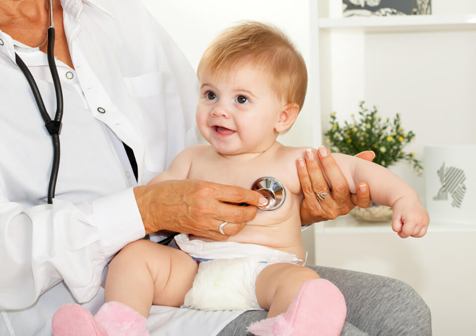
You Can Listen To Your Baby’s Heartbeat With The Stethoscope!
For 12 weeks, when the fetal bone marrow begins to produce blood cells, stimulating blood circulation continues.
Following 17 weeks, the fetal cerebrum starts to control the heartbeat in planning to help the child to get involved step by step in the outside world.
(So far, the heart was pulsating unexpectedly.) After three weeks; around 20 weeks; then you can hear the child’s pulse with a stethoscope.
If your doctor needs better listening (and vision); we recommend an echocardiogram of the fetus from 18 to 24 weeks; which is a special ultrasound to assess the fetal heart (if you have a family history of congenital heart disease; or if you have diabetes, phenylketonuria; or an autoimmune disease, make sure you have).
At this time, the child’s heart beats with about 140 beats. At the conclusion of the week; 25 capillaries (the littlest blood vessels) are shaped and filled with blood.
Capillaries allow the blood of oxygen through the arteries of the heart to move to the tissues of the baby’s body as a whole; making these small vessels the central component of the circulatory system.
The Heart Of The Baby At Birth
The circulatory system of the baby will continue to grow slowly and steadily, so it will be ready to debut outside the uterus at 40 weeks.
Whereas the fetal circulatory framework creates quickly amid pregnancy; it really works very in an unexpected way within the womb than it does after birth.
Some time recently births, the baby’s lungs still don’t work; because the infant does not breathe within the womb.
Until your child is born and takes those, to begin with; free breaths, your creating circulatory framework is for consistent blood supply; wealthy in oxygen and supplements the supply routes and umbilical veins carry the unoxidized blood and squander to be expelled and after that the child needs from you.
Some other differences: in the fetal heart there is a shunt or a label that removes blood from the lungs (simply because it is not needed in the uterus).
Like you, your kid has an aspiratory conduit (which conveys blood from the heart to the lungs) and aorta (which conveys blood from the heart to the body).
Be that as it may, they are associated with another vessel (blood vessel channel); which additionally serves to occupy blood from the lungs in the uterus.
At long last, your little one has an opening in the uterus just during the upper cardiovascular hole (porous oval structure); which again redirects blood from the lungs.
As soon as the baby is born, the difference between these fetuses disappears completely (or begins to disappear).
At the point when the umbilical rope is cut, the child’s lungs enter the air; the fetal course framework closes down; and the shunt starts to close. All systems go for children.
How To Keep The Child’s Heart-Healthy
It creates and changes especially when your kid is in the belly.
Some things, such as genetic abnormalities; may affect the development of your child’s mind are out of your control; but your child’s tinnitus may be a symptom of an underlying disease:
Taking folic acid before pregnancy and during pregnancy helps to prevent congenital heart disease in infants.
In case you’re a smoker; stop it as quickly as time permits: the specialists found that smoking moms in the primary trimester; can represent up to 2 percent of all heart deserts.
In the event that you have Type 2 diabetes or gestational diabetes; it is essential to screen your glucose levels leveled out during pregnancy since diabetes is related to an expanded danger of heart deserts.
Not Acutance (acne), the fetus is the heart.
Alcohol.
Regardless of whether you play it safe and do everything suggested by your primary care physician; you might be brought into the world with inherent heart abandons.
There are many factors that are not your fault; Most of which are out of your control; that can cause heart defects, and the doctor just News is that early detection and your child will be able to get the treatment he needs to increase the possibility of sending a long and healthy life.
Read Also:
- How to keep baby’s heart to be healthy during pregnancy?
- Here You Will Get the Cute baby Girl Games.
- Healthy Pregnancy – Is It Safe to Drink Soy Milk During Pregnancy?