A pregnancy ultrasound is a study that utilizes high-frequency sound waves to picture the growing child as well as the reproductive organs of the mother. The average amount of ultrasounds depends on the pregnancy.
An ultrasound, also known as a sonogram, can assist in monitoring average growth and tracking future issues. In addition to conventional ultrasound, there are several more sophisticated ultrasounds— including a 3-D ultrasound, 4-D ultrasound, and fetal echocardiography that shows the core of the fetus in detail.
What are the uses of ultrasound?
Ultrasound may be used at different times during pregnancy, including:
- First-quarter ultrasound
Used to test for the development of the embryo in the womb, confirm the number of embryos, and compute the gestational age and the baby’s due date.
- Second trimester
Ultrasound between the 18th and 20th weeks is used to verify fetal constructions such as the spinal tract, teeth, body, and inner organ growth. The placenta size and place are also confirmed. If the family wants to learn, the sex of the baby can be checked.
- Third trimester
After 30 weeks, ultrasound is used to verify that the child continues to develop at a standard pace. The position of the placenta is inspected to ensure that the cervix is not blocked.
What occurs in an ultrasound session?
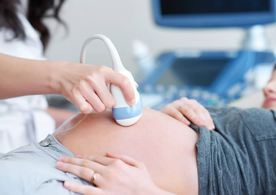
During the exam, you are on an observed exam chair, and a small number of water-soluble gel is added to your skin over your stomach. The gel will not harm your flesh or your garments.
A tiny device is softly pushed against the skin of your belly, called a transducer. The transducer transmits sound waves of high frequency into the body, which represent inner buildings, including your child. The sensor receives the sound waves or reflecting echoes and converts into an image on display. These images can be printed or recorded in a videotape sometimes.
During the exam, there is nearly no trouble. If the experiment requires a full bladder, you may experience some pain when the sensor is placed over the bladder.
No specific arrangement for the ultrasound test is prepared. Some consultants ask you to take 4-6 glasses of water before the analysis, so you have a full bladder. It helps the doctor to see the baby on the ultrasound better. You will be requested to avoid urination until after the test.
Some doctors permit you to record the ultrasound and take it home. Inquire to your doctor if this is an option. If this is the case, you must bring a blank videotape or DVD to your meeting.
Read Also:
Why should I have an ultrasound on my third month of pregnancy?

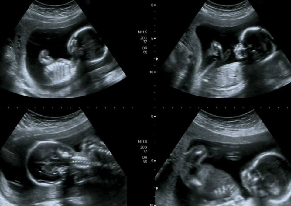
Your baby weighs approximately 23 grams (0.8 oz) at 13 weeks and is about 7.4 cm (2,9 in) long from crown to rump, around the length of a pea pod. All its organs and muscles are in place, although they still have a lot to do. At 3 months of pregnancy, ultrasound can give you a glimpse of your baby.
This month, the facial muscles of your baby train, as their small traits form one expression after the next. She/He is going to be squinting and grimacing, and sometimes she/he even has hiccups when she practices breathing movements. Their body is developing a fine coating of hair, called lanugo, which helps them to keep their bodies warm. Before birth, most babies shed this hair.
Your baby’s brain also develops the part responsible for memory and problem-solving. Her/His heart is now entirely shaped and is three times faster than yours. She/He increases and will have reached approximately 11.6 cm (4.6 in) by week 16 about the size of an avocado. She/He’s even going to start having small fingernails.
What kind of ultrasound should I get?
More advanced techniques of ultrasound can be used when a more detailed image is required. You may provide the doctor with the information needed to make a diagnosis if your traditional ultrasound has detected problems.
Transvaginal
Transvaginal ultrasound can be performed to create a clearer picture. This ultrasound is more likely to be used in the early stages of pregnancy when a clear picture is more challenging to capture. A small ultrasound test is inserted in the vagina for this test. The analysis rests on your vagina’s back while the images are being achieved.
3-D
In contrast to traditional 2-D ultrasounds, a 3-D ultrasound can show the width, height, and depth of your fetuses and organ to your doctor. This ultrasound can help you, in particular, to diagnose suspected problems during pregnancy.
The process follows the same as the standard ultrasound, but a particular 3-D image sample and software are used. Specialized training is also required for the technician to prevent it from being as widely available.
4-D
A 4-D ultrasound can also be referred to as a dynamic 3-D ultrasound. A 4-D ultrasound, unlike other ultrasounds, produces a moving fetal video. It creates a better image of the face and motions of the baby.
It also better captures shades and highlights. Similar to other ultrasounds, this ultrasound is carried out with special equipment.
Echocardiography
Fetal echocardiography is performed if your doctor suspects that your baby has heart defects. This test may be done similarly to a traditional ultrasound for pregnancy, but it may take longer. It captures a thorough image of the heart of the fetus–one that shows the size, the form, and the structure of the heart.
This ultrasound also gives your doctor a look at the functioning of your baby’s heart, which can help diagnose heart problems.
Is it safe?
Ultrasound is a secure, painless, non-invasive treatment. Many parents view the ultrasound as a chance to see and perhaps learn about their unborn baby. You should acknowledge, nevertheless, that the ultrasound is an indicative method and may propose, in some cases, that a fetus is abnormal. Additional exams are generally necessary to verify the diagnosis.
Takeaway
What occurs next relies on your ultrasound outcomes. Note that a typical result will not guarantee that your baby is healthy, as this test can not identify any defects. If fetal abnormalities have been identified, further testing may be needed to verify the diagnosis.
These tests are optional, including amniocentesis and sampling of the chorionic villus. Talk to your doctor about the advantages, uncertainties, and complexities of these examinations before determining to proceed or not.
Read Also:
- A Guide To The Rare Cases of Cryptic Pregnancies
- Baby Not Growing in the Womb How To Deal With It
- How to Keep Baby’s Heart to be Healthy During Pregnancy?