
Every parent is excited to see and observe what is happening to their babies inside their mothers’ womb. Thanks to modern technology, it is possible to have a glimpse of your unborn baby through ultrasound. Nothing is more satisfying to expectant parents than seeing their babies grow and develop.
Pregnant mothers should have regular check-ups and trips to their doctors. It will assure the safety of both the mother and the baby. Today, it is a vital routine for pregnant women in their second trimester to undergo the 20-week anatomy scan. What is it? And what is this for?
Read also: 20 Weeks Pregnant
What is a 20-Week Anatomy Scan?
The 20-Week Anatomy Scan is also known as the Level 2 Ultrasound. As a pregnant woman approaches her 20th week or the second trimester, this special pregnancy ultrasound is required to see how the baby is developing. The session may take about 30 to 45 minutes, depending on your baby’s cooperation. Most parents take home a photo of their babies from this session to start their babies’ photo album and milestone journals. the Level 1 sonogram on the first trimester confirms the date of pregnancy, while the Level 2 sonogram is a more detailed scan. It can give additional information and updates about babies’ growth inside the womb, how it’s going on and what may lie ahead of your pregnancy and after it.
Read also: Non-Growing Fetus: Symptoms to Check
How is it done?

An expectant mother is required to drink water to have a full bladder before the 20-week ultrasound. It makes taking ultrasound images easier. She will recline in the examination table with her tummy exposed. The sonographer will apply the gel to the tummy and will move a gadget that looks like a wand, the transducer over the abdomen. The computer will then convert the sound to 2D, 3D or even 4D images.
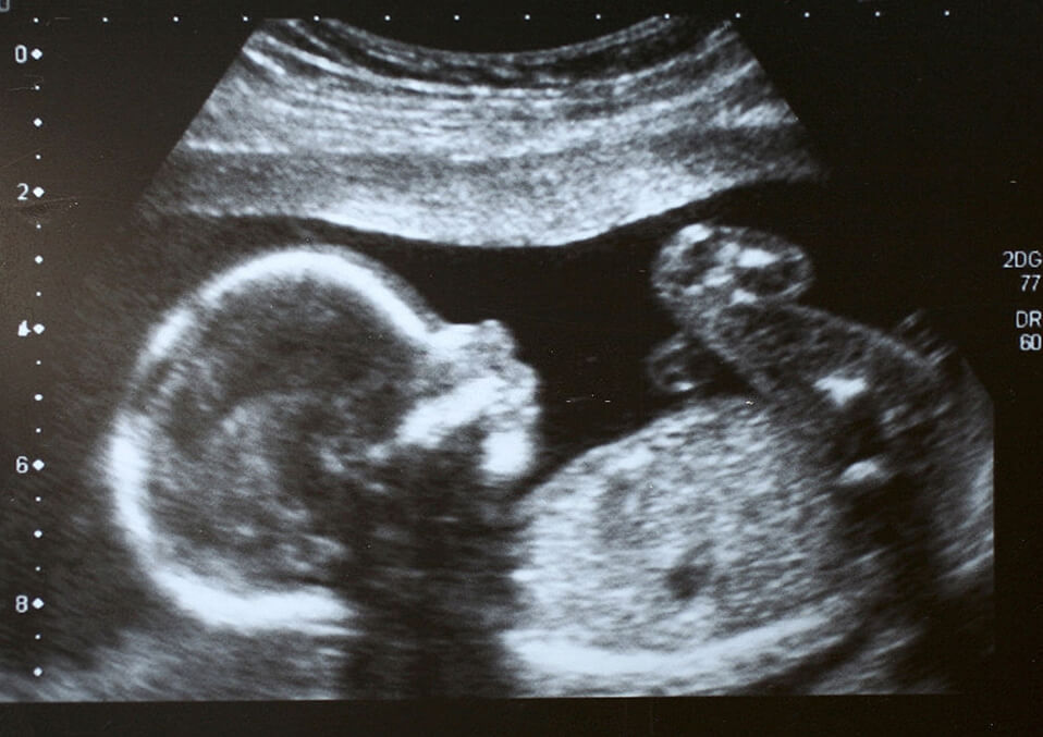
For a more accurate assessment, the sonographer will be getting different views from different angles. Once he gets a clear shot, he will pause or freeze-frame the image. This is the actual sonogram and this is a clearer image than level 1 ultrasound. In the scan, you can know the size of a specific part of the baby’s body. You can see your baby’s beating heart, the curve of the arms, the legs, the face, and the spine. There are times when you can even capture your baby while sucking his or her thumb.
What is this for?
The 20-week anatomy scan focuses on the anatomy of the fetus to assure that everything is doing okay and the baby is developing or growing as it should. With the 20-week ultrasound, baby’s measurements will be taken. The baby will be measured from head to foot, the middle, the head, and the baby’s weight. It can check on the baby’s heart, kidneys, bladder, stomach, spine, brain, and the sex organ. The amniotic fluid, the placenta, and the baby’s heart rate and position will also be checked.
This process can check and detect possible abnormalities in the baby early. Additional testing can be done once the need arises.
What are the risks?
As of now, there are no dangers associated with the 20-week anatomy scan. Practice necessary precautions and avoid too much exposure to the ultrasound during your pregnancy. Worry not, your practitioner will interpret and explain to you the images on the computer while doing the procedures
A final note
The 20-week anatomy scan is such an exciting milestone for parents as they can take a peek of their baby with a clearer image. Do it with your partner for a more special and memorable experience and surely you’ll both fall in love with your child at first, clear sight.
Read also:
- Ectopic Pregnancy: Ultrasound Diagnosis
- What To Expect On The First Ultrasound: 12 Most Important
- What Should You Know When Doing the Ultrasound at 20 Weeks of Pregnancy
